Taurodontism: Understanding This Unusual Dental Condition
TAURODONTISM
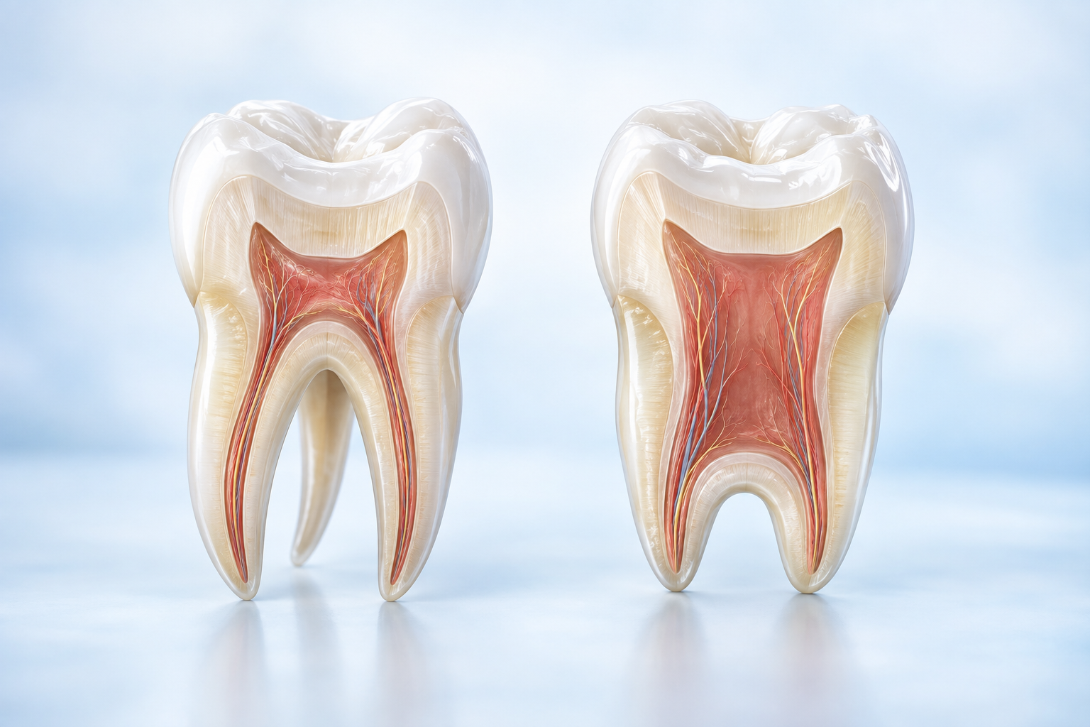
Taurodontism is a dental condition where the tooth shape changes because a part of its development, called Hertwig's epithelial sheath diaphragm, does not fold in at the right level.
Taurodontism is a condition where the inside part of a tooth (the pulp chamber) is larger than usual. The floor of this chamber is positioned lower than normal, and the area where the crown and root meet (the cemento-enamel junction) doesn’t have the usual narrowing. It mostly affects adult molars but can also occur in baby teeth. It can appear on one side or both sides of the mouth and may affect different teeth or sections of the mouth in various ways.
Etiology of Taurodontism
Taurodontism can be caused by a variety of factors. These include:
- Hormonal disorders or mutations: Hormonal imbalances or disruptions during tooth development can lead to the morphological changes observed in taurodontism.
- Genetic disorders or mutations: Specific genetic conditions, such as amelogenesis imperfecta (a disorder affecting enamel formation) and ectodermal dysplasia (a condition involving abnormal development of the skin, hair, nails, teeth, and sweat glands), have been linked to taurodontism. These disorders interfere with the normal developmental processes of the teeth, resulting in variations in shape and structure.
Clinical and Radiographic Features
Taurodontism predominantly affects molars, and understanding the development of these teeth is essential for the diagnosis and treatment of the condition. On radiographic examination, taurodontism is identifiable by an increased pulp chamber (inside part of the tooth) size and a lack of constriction at the level of the cemento-enamel junction. Cone beam computed tomography (CBCT) imaging which is a special type of 3D radiograph is particularly useful for diagnosing taurodontism, as it provides high-resolution images that allow clinicians to visualise the altered tooth shape and size in greater detail. Routine radiographs is especially important for patients with genetic disorders, as taurodontism may not be evident during a routine oral examination.
Taurodontism has been associated with several developmental syndromes and abnormalities, further complicating its clinical management. These include:
- Down syndrome
- Klinefelter syndrome
- Mohr syndrome
- Wolf-Hirschhorn syndrome
- Lowe syndrome.
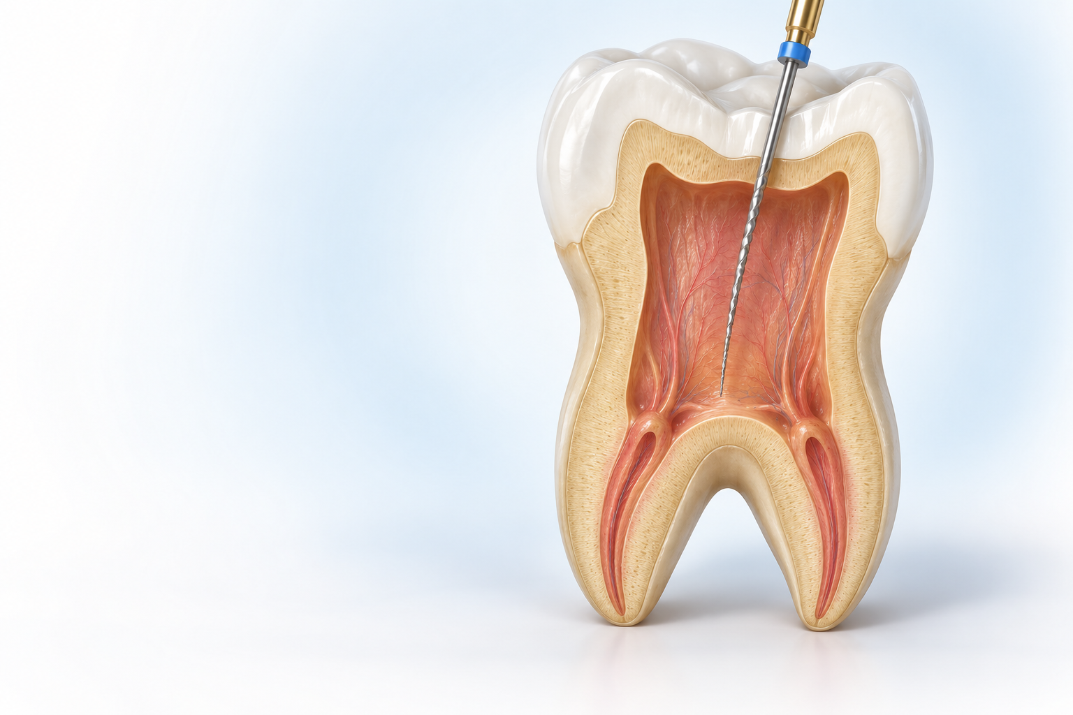
The management of taurodontic teeth presents several challenges for dental practitioners, particularly in the areas of pulp therapy and tooth extraction. Taurodontic teeth have a larger inner chamber (pulp chamber) that can vary in size and may contain hard deposits called pulp stones. This can make root canal treatment more challenging because the openings to the root canals are lower than usual, and the larger pulp can cause more bleeding during procedures. Additionally, since the roots are shorter and the chamber floor is lower, it can be harder to fully clean out dead tissue during treatment. When doing a root canal on taurodontic teeth, dentists need to be extra careful to avoid accidentally drilling through the tooth or root. This is because these teeth have an unusual shape, making the procedure trickier than usual. Additionally, extracting a taurodontic tooth is often more challenging than extracting a normal tooth, as the dilated roots in the apical third of the tooth complicate the procedure. Specialised techniques and equipment may be required to ensure safe and effective extraction, if necessary.
In conclusion, taurodontism is a significant dental anomaly that requires careful consideration for diagnosis and treatment. Although it is most commonly observed in permanent molars, it can affect both permanent and deciduous (baby teeth) teeth, and it may be associated with a variety of genetic syndromes and abnormalities.